Anatomy, histology and physiology
The esophagus is a muscular tube that connects the throat
(pharynx) to the stomach. It is approximately 25 cm (10 inches)
long in adults and is located behind the trachea (windpipe) and
in front of the spine. The esophagus is divided into three
parts: the cervical esophagus (the upper part), the thoracic
esophagus (the middle part), and the abdominal esophagus (the
lower part that connects to the stomach). The esophagus is lined
with a mucous membrane that produces mucus to help lubricate
food as it passes through. The walls of the esophagus contain
several layers of muscle that contract in a coordinated manner
to propel food from the throat to the stomach through a process
called peristalsis. At the lower end of the esophagus, there is
a ring of muscle called the lower esophageal sphincter (LES)
that helps prevent stomach acid and contents from flowing back
up into the esophagus (acid reflux).
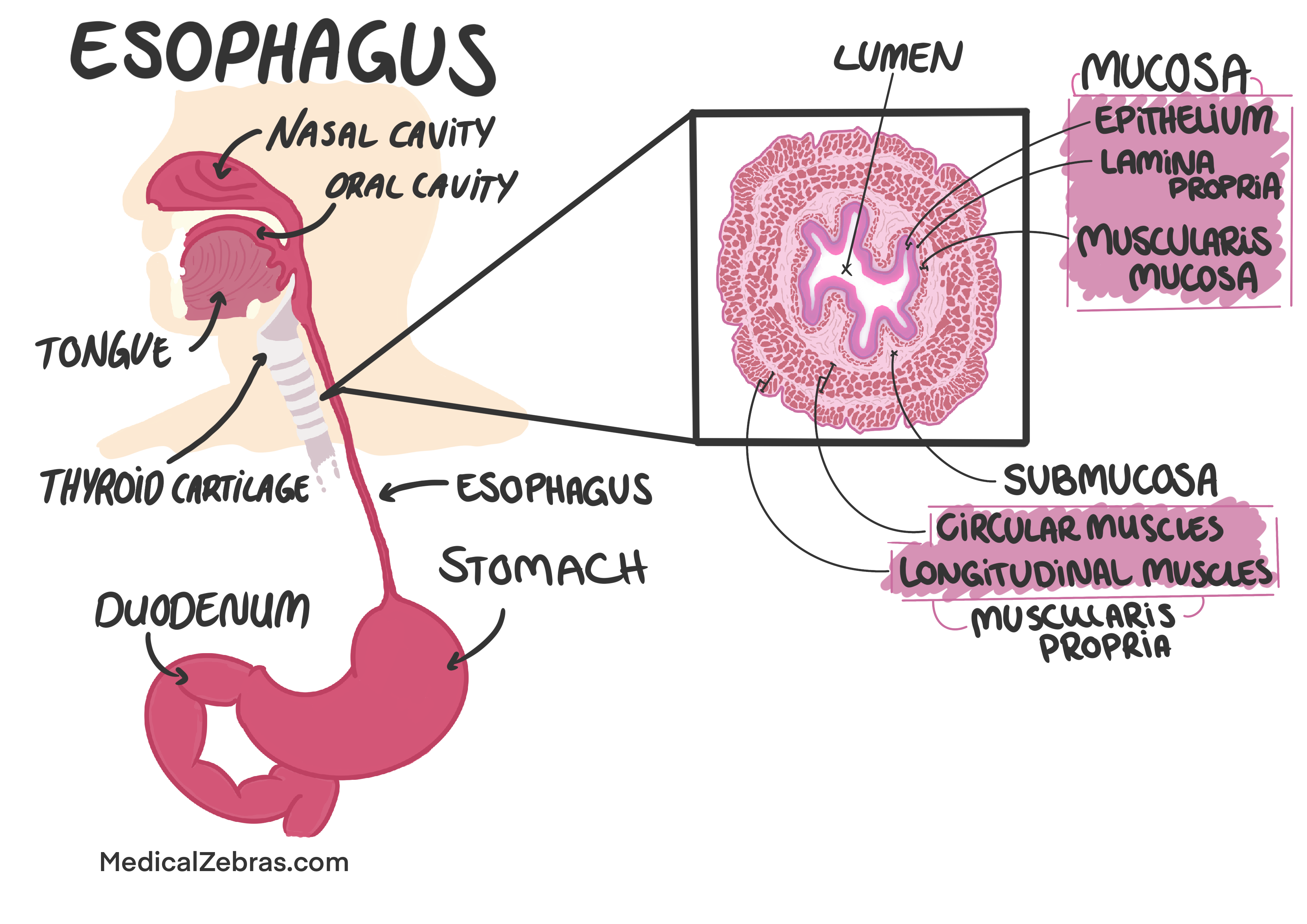
Histology
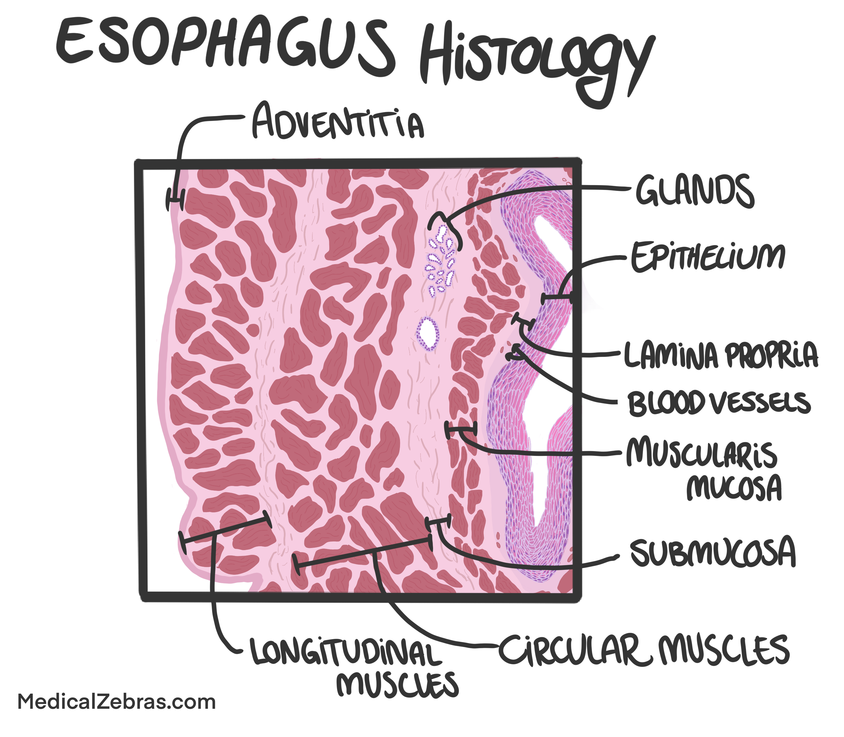
The histology of the esophagus consists of several layers:
-
Mucosa: The innermost layer, which is lined with stratified
squamous epithelium that protects against mechanical stress
from food passage.
-
Epithelium: Stratified squamous epithelium that provides a
protective barrier.
Lamina propria: A layer of connective tissue that contains blood vessels, lymphatics, and immune cells.
Muscularis mucosae: A thin layer of smooth muscle that helps with local movements of the mucosa.
-
Submucosa: A layer of connective tissue that contains blood vessels,
nerves, and glands that produce mucus to lubricate the
esophagus.
-
Muscularis externa: A layer of muscle responsible for peristalsis. The upper
third of the esophagus contains skeletal muscle, the middle
third contains a mix of skeletal and smooth muscle, and the
lower third contains smooth muscle.
-
Adventitia: The outermost layer, which is made up of connective
tissue that anchors the esophagus to surrounding structures.