Arteries & Veins
Arteries
Arteries are blood vessels that carry oxygenated blood away from the heart to the tissues of the body. They have thick, muscular walls that can withstand the high pressure of blood being pumped from the heart.Histology
-
Tunica intima
the inner layer of the artery, composed of endothelial cells,
underlying loose connective tissue and internal elastic lamina
(elastic membrane) that separates tunica intima from tunica media.
The endothelial cells tend to proliferate as response to injury
(altered blood flow or increased pressure). This layer is affected
by progressive intimal fiborsis, that is especially prominent in
the thyroid, spleen and myometrium.
-
Tunica media
the middle layer of the artery, composed of vascular smooth muscle
cells, elastic tissue and collagen.
-
Tunica adventitia
it the outer most layer, composed mostly of collagen fibers,
elastic fibers and fibroblasts. You might also see macrophages and
other inflammatory cells, ganglion cells and vasa vasorum (small
blood vessels that supply the larger ones).
Types of arteries
-
Elastic arteries
are the largest arteries in the body (for example the aorta and
its major branches). They have a large amount of elastic fibers in
the tunica media, which allows them to stretch during systole and
recoil during diastole, helping to maintain a continuous blood
flow throughout the cardiac cycle.
-
Muscular arteries
are medium-sized arteries that have a higher proportion of smooth
muscle cells in the tunica media compared to elastic fibers. This
allows them to regulate blood flow to specific organs and tissues
by constricting or dilating.
-
Arterioles
are the smallest arteries that lead to capillaries. They have a
thin tunica media with only a few layers of smooth muscle cells.
Arterioles play a crucial role in regulating blood flow and blood
pressure by constricting or dilating in response to various
stimuli.

Veins
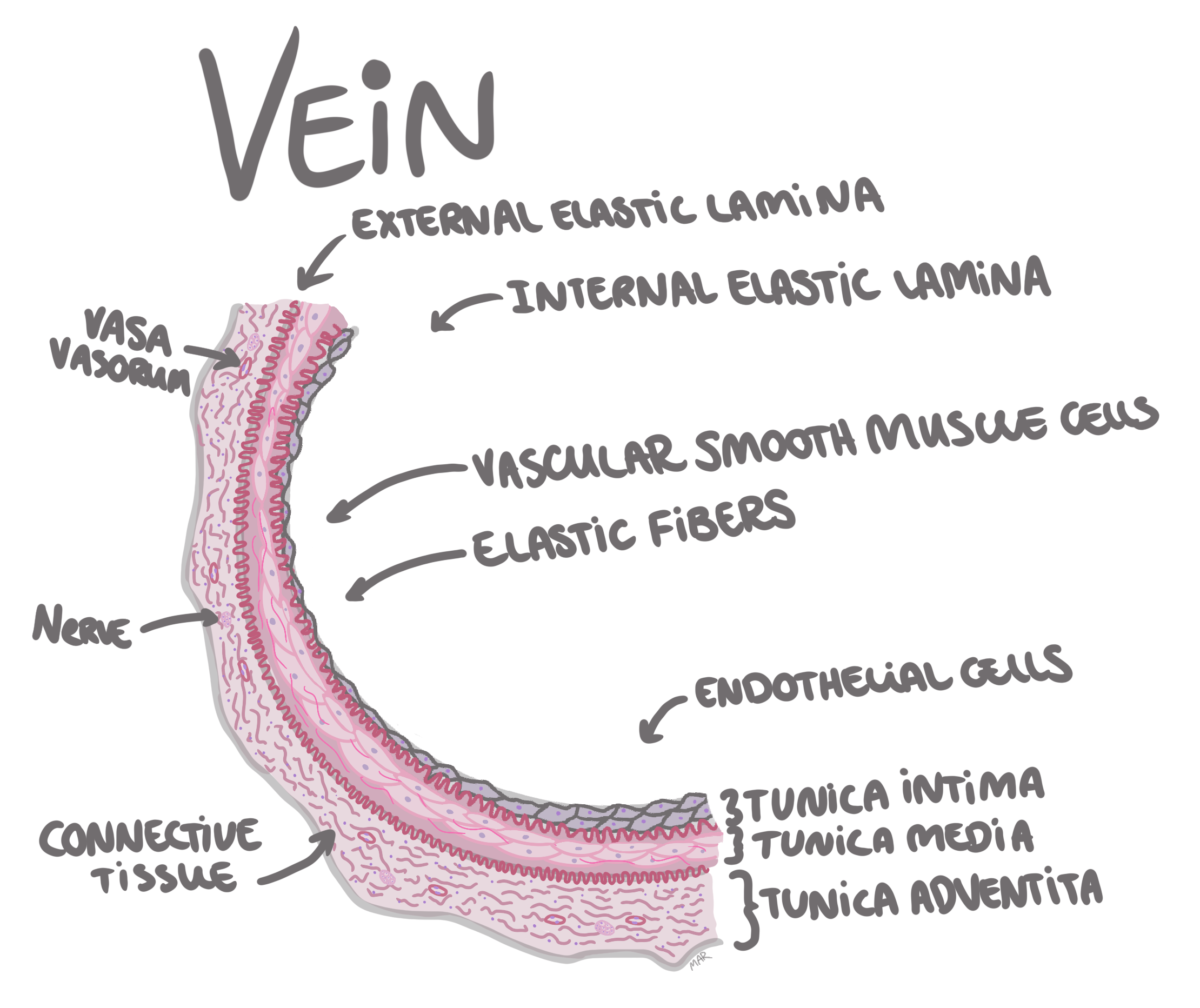
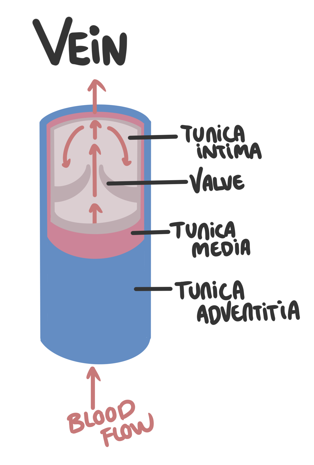
Veins are blood vessels that carry deoxygenated blood from the tissues back to the heart. They have thinner walls than arteries and often have valves to prevent backflow of blood.Histology
-
Tunica intima
the inner layer of the vein, composed of endothelial cells,
underlying loose connective tissue and internal elastic lamina
(elastic membrane) that separates tunica intima from tunica media.
-
Valves: Because the veins are responsible for continuous bloodflow
back to the heart the tunica intima makes extra folds now and
then to form, most often, bicuspid valves (2 flap valve). These
valves make continuous bloodflow to the heart possible by
hindering backflow (the valves close). If the veins are dilated
(varicose veins) or the valves are damaged (for example
post-thrombotic syndrome), the valves will not close properly
and backflow can occur, leading to venous insufficiency. These
valves are more common in the extremities, where the blood has
to fight gravity to get back to the heart.
-
Tunica media
the middle layer of the vein, composed of vascular smooth muscle
cells, elastic tissue and collagen. This layer is way thinner in
veins, as veins are not exposed to the same high pressure as
arteries.
-
Tunica adventitia
it the outer most layer, composed mostly of collagen fibers,
elastic fibers and fibroblasts. You might also see macrophages and
other inflammatory cells, ganglion cells and vasa vasorum (small
blood vessels that supply the larger ones).
Types of veins
-
Superficial veins
are located close to the surface of the skin and are responsible
for draining blood from the skin and subcutaneous tissues. They
are often visible through the skin and can be affected by varicose
veins.
-
Deep veins
are located deeper in the body and are responsible for draining
blood from the muscles and organs. They are larger and have a
higher capacity than superficial veins.
-
Venules
are the smallest veins that receive blood from capillaries. They
have thin walls and a small amount of smooth muscle cells in the
tunica media.
Pathology
Atherosclerosis
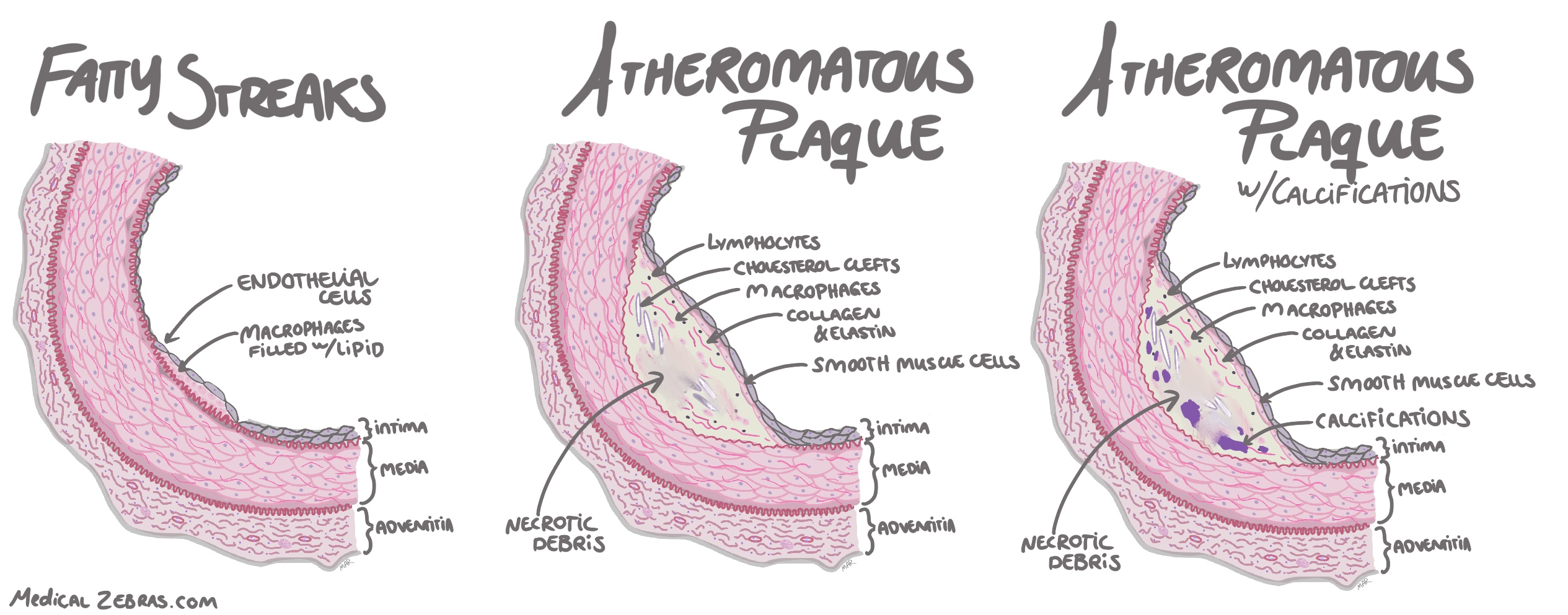
Hypertension and blood flow turbulance can cause endothelial/vascular damage, which in turn leads to increased endothelial permeability, leading to leukocyte adhesion and accumulation of lipids in the intima, and with it proliferation of smooth muscle cells. Leading to a thickened intima. This is the first step in atherosclerosis.One of the most common place for artherosclerosis is where the arteries bifurcate (split), as the bifurcation cases blood flow turbulance.
-
Fatty streaks:
a visible yellow to white lesion on the intimal surface, where
macrophages filled with lipid have accumulated beneath the endothelium
in the intima.
-
Atheromatous plaques:
are raised yellow to white lesions within the intima. The lesion
contains a lipid core and necrotic debris covered by a fibrous cap.
The fibrous cap contains smooth muscle cells, macrophages filled with
lipid, collagen and elastin as well as lymphocytes. The necrotic
center contains cellular debris, macrophages filled with lipid,
cholesterol crystals and calcium. This plaque can increase in size
with time and therefore protrude into the lumen causing stenosis
(decreased lumen) and affect the quality of the media underneath which
in some cases can lead to aneurysm formation.
-
Calcification:
As the lipid core in the atheromatous plaque contains calcium, this
accumulation with time will lead to calcifications within the plaque.
Calcium mainly comes from dying cells that leak calcium into the
extracellular matrix.
-
Stenosis:
is when the atheromatous plaque protrudes into the arterial lumen
decreasing the diameter of the lumen, thus disturbing and decreasing
blood flow.